Unlocking the Mystery of Microtubule Growth: The Intricate Dance of Kip2 and Bik1
In a multidisciplinary study newly published in the Journal of Cell Biology, the Barral group (ETHZ, D-BIOL/IBC) together with the Steinmetz group (PSI), the Stelling group (ETHZ, D-BSSE) and the Liakopoulos lab (CNRS, Montpellier) sheds light on how the motor domain of the kinesin protein Kip2 collaborates with the microtubule plus-end-binding protein Bik1, to moonlight as a microtubule polymerase.

Kinesins are microtubule-dependent motor proteins that are best known for their role in transporting cargos along microtubules. The findings now published in JCB change our understanding of how some of these motor proteins can also control the organization of the microtubule cytoskeleton. The authors show how Kip2, a cytoplasmic kinesin, promotes microtubule growth and stabilization. Previous studies had shown that cells lacking the KIP2 gene form shorter and less abundant astral microtubules. Intriguingly, Kip2's microtubule-stabilizing function in vivo depends on the presence of the cytoplasmic linker protein Bik1, the yeast orthologue of metazoans' CLIP-170. The new study indicates that two critical structural elements underlie the ability of Kip2 to promote microtubule polymerization: an interaction interface in the motor domain dedicated to binding free tubulin, in addition to the microtubule shaft, and a binding site for the cytoplasmic linker protein Bik1 in its C-terminal tail. While the free tubulin-interaction interface is not involved in Kip2's motility along microtubules, it plays a crucial role in microtubule polymerization. The binding site for Bik1, on the other hand, increases Kip2's residence time at microtubule plus-ends in living cells, in a Bik1-dependent manner.
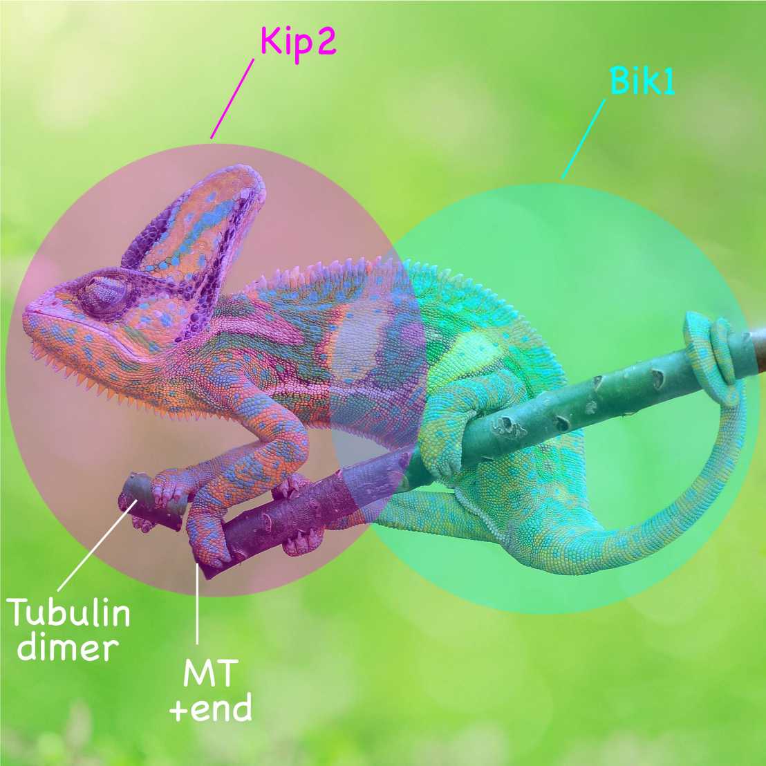
Based on their findings, the researchers have now developed a model for how Kip2 contributes to microtubule polymerization in vivo. They suggest that when Kip2 reaches the very end of a microtubule through its motile translocation, the free motor domain of Kip2 binds free tubulins and promotes its incorporation into the protofilament. In other words, Kip2 might extend its own track beneath its "feet" once it arrives at microtubule tips. Although more detailed mechanistic studies will be needed to address this possibility, the position of the free-tubulin-binding interface in the motor domain suggests that the delivery and release of tubulin dimers at the elongating microtubule end is ruled by the same mechanisms as those mediating binding and release of tubulin on the shaft during motility, i.e., depend on the ATPase cycle of Kip2. This idea is consistent with previous observations, indicating that ATP hydrolysis is essential for Kip2 to promote microtubule growth.
Link to the paper in external page "Journal of Cell Biology"